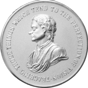
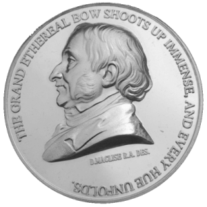
Newton Medal 2024 Lecture: ‘Variability, colour thresholds and chromatic mechanisms’
Abstract
It is not unreasonable to expect some differences in human visual perception given the innumerable differences between human eyes, and the role inferential brain mechanisms play in generating a representation of what is most likely to be present in the visual scene. Many inter subject differences remain poorly understood, but the advantages of ‘seeing’ things ‘the same way’, often makes us forget about the real differences.
Fortunately, some limits are imposed on what we all see by the largely invariant properties of the five, spectrally distinct sensors in the eye. What we know for sure is that much of the spatial information carried in modulations of intensity and spectral content in retinal images is captured in four, retinal photoreceptor pigments. This information is then ‘condensed’ in the retina to provide us with efficient encoding of edges and contours defined by either luminance contrast or by spatial variations in the spectral content of the image. The latter form the basis for the red / green and yellow / blue chromatic signals. Other mechanisms which operate best at lower light levels and rely on both excitatory and inhibitory interaction of signals from rods and cones to make it all work, enable the human eye to function over an enviable range of light levels. In addition, the output of a small population of ganglion cells with large receptive fields, which receive inputs from virtually all photoceptors, is also modulated by melanopsin, a fifth photopigment located within the cell, which responds at higher light levels. These ganglion cells form a separate vision channel which may play an important role in sensing the amount of light in the visual scene and may also contribute to our ability to judge the level of ambient light. The effectiveness of these different signals generated in the retina varies greatly with changes in stimulus conditions, but all these signals can contribute directly and can affect the appearance of the objects we see.
In this lecture I shall make use mostly of colour vision and shall focus on how either changes in or the absence of one chromatic mechanism can influence the effectiveness of the remaining vision channels and how this can affect our functional vision and the ability to carry out visual tasks, including those involved in colour assessment.
In order to understand the results of colour assessment tests when plotted in colour spaces defined for subjects with normal colour vision, we developed a new model of colour discrimination to derive predictions of colour thresholds in subjects with variant cone pigments and to describe how photopic and scotopic luminance signals can affect colour assessment outcomes under conditions that are deemed to be photopically isoluminant for subjects with normal colour vision. The model also predicts accurately how the selective absorption of short wavelength light by the lens and the macular pigment in the eye can affect the orientation and size of colour threshold ellipses. Equally important, a default outcome of the model is that colour threshold contours can be plotted as circles to produce a uniform colour space. When the combined colour threshold signal strength needed to just see a colour difference between the stimulus and its adjacent background is no longer invariant with the direction of chromatic change, the results are indicative of either congenital or acquired colour deficiency.
In addition to normal colour vision and congenital colour deficiency, I shall also be presenting results from clinical studies to illustrate how functional vision can change when the binocular summation and inhibition of signals no longer functions normally or when the brightness channel no longer signals the presence of light.